Technology
Microscope-Guided Precision Dentistry
A dental operating microscope is a stand-mounted scope with its own bright light, positioned over your tooth while we work. It magnifies far beyond what loupes or the naked eye can show, which is what makes a hairline crack, decay under an old filling, or the inside of a root canal checkable as we go.
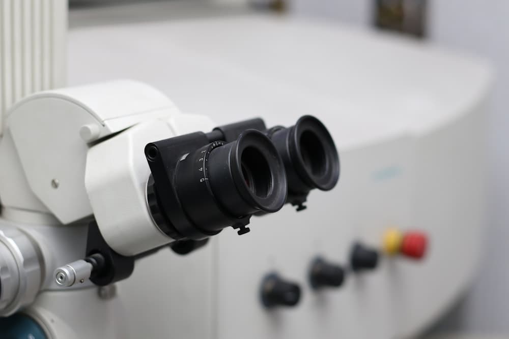
A dental operating microscope gives bright, magnified views of a tooth inside and out, well past what loupes or the naked eye can reach. That level of detail counts most in work where a fraction of a millimeter decides whether a tooth is saved or lost.
From your side, the appointment looks much like any other. You lie back and we position the scope above you. It never touches you and nothing extra goes in your mouth. The light is bright, so we may hand you glasses or ask you to keep your eyes closed. We also work in silence for stretches, because looking carefully takes concentration.
On a root canal, magnification is used at every stage. Finding the canals. Cleaning each one to the end. Checking the walls for a channel that is easy to miss. Confirming the filling material has gone where it should. The scope's own light is as much the point as the magnification, because the entrance to a canal reads as a shadow until something is shining straight down it.
In an exam, the scope shows a crack too fine to see without magnification. A hairline fracture in a back tooth can explain pain on chewing that nothing else accounted for. Decay creeping under the edge of an old filling shows up the same way, while it is still small and the repair is still simple.
Magnification also changes how much tooth we take away. When you can see the exact border between sound tooth and decay, you can stop at that border. Less drilling leaves more of your own tooth, and your own tooth is the part worth keeping.
The alternative is loupes, or no magnification at all, plus experience and feel. Careful dentists do good work that way. The difference is verification. Under the scope, each step is looked at as it happens rather than assumed to have gone to plan.
Magnification is not useful everywhere, and we do not use it for everything. A cleaning or a straightforward filling does not call for it. It can add time to an appointment, because careful work is slower work. And finding a problem early does not always mean the tooth can be saved. When a crack runs too far, we explain what we are seeing and give you the honest options.
Benefits
Why it matters for you
- Finds fine cracks and hidden decay while they are still small
- Shows the border between sound tooth and decay, so less tooth is removed
- Every step of a root canal is checked as it happens
- Helps locate canals that are narrow, curved, or easy to miss
- Existing crowns and fillings can be inspected at the margin
- We explain what we find before you decide anything
Where We Use It
Procedures it supports
- Root canal therapy
- Detecting cracked teeth
- Conservative cavity preparation
- Evaluating and refining existing restorations
- Retreating a root canal that did not settle
- Locating canals in a tooth with unusual anatomy
Questions
Common questions
No. We use magnification where it meaningfully improves what we can see and do, mainly root canals and detail-critical restorative work. A cleaning or a straightforward filling does not need it. You will know when it is part of your treatment, because we tell you what we are doing and why.
It can. Careful work is slower work, and checking each step takes time the step itself does not. On a root canal, that is time we consider well spent. We will tell you at the start how long to plan for, so the visit does not run past what you expected.
No. The scope sits above you on a stand and never touches you, and nothing extra goes in your mouth. What you notice is the light, which is bright, and the quiet while we work. The treatment itself feels like the same procedure, done at a more careful pace.
The scope puts magnification and a direct beam of light down the access opening, which is what makes the inside of a root visible. Under the scope we can find the canals, clean each one to the end, and look for a channel that is easy to miss. Working from what we can see beats working from feel.
Sometimes seeing the detail changes the plan, and sometimes it does not. A crack found early can often be restored, while one running deep into the root may not be. We explain what we are seeing and tell you honestly which of those we think you have.
See it for yourself