Smile Gallery
Before and after, from our own patients
Every photo on this page is an Avora Dental patient treated by Dr. Aashil Shah. No stock images, no stand-ins. Drag the slider on each case to move between the before photo and the after photo of the same patient. Each one is a single person with a single set of teeth, and what worked for them may not be the right plan for you.
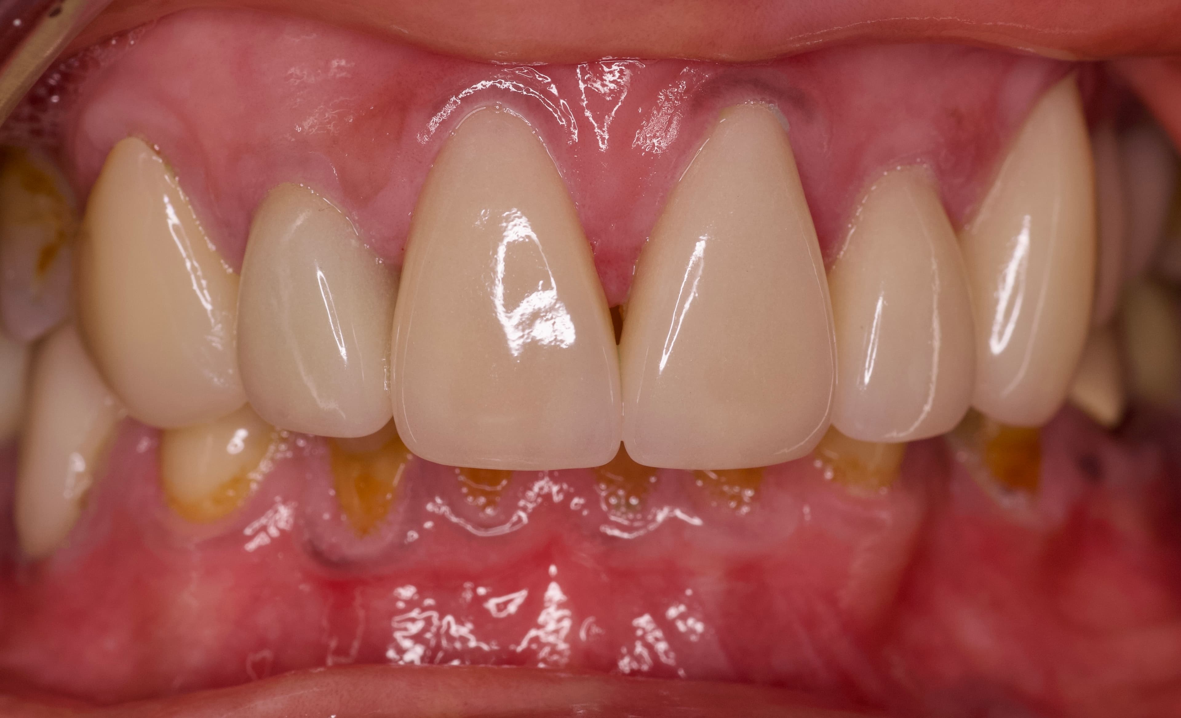
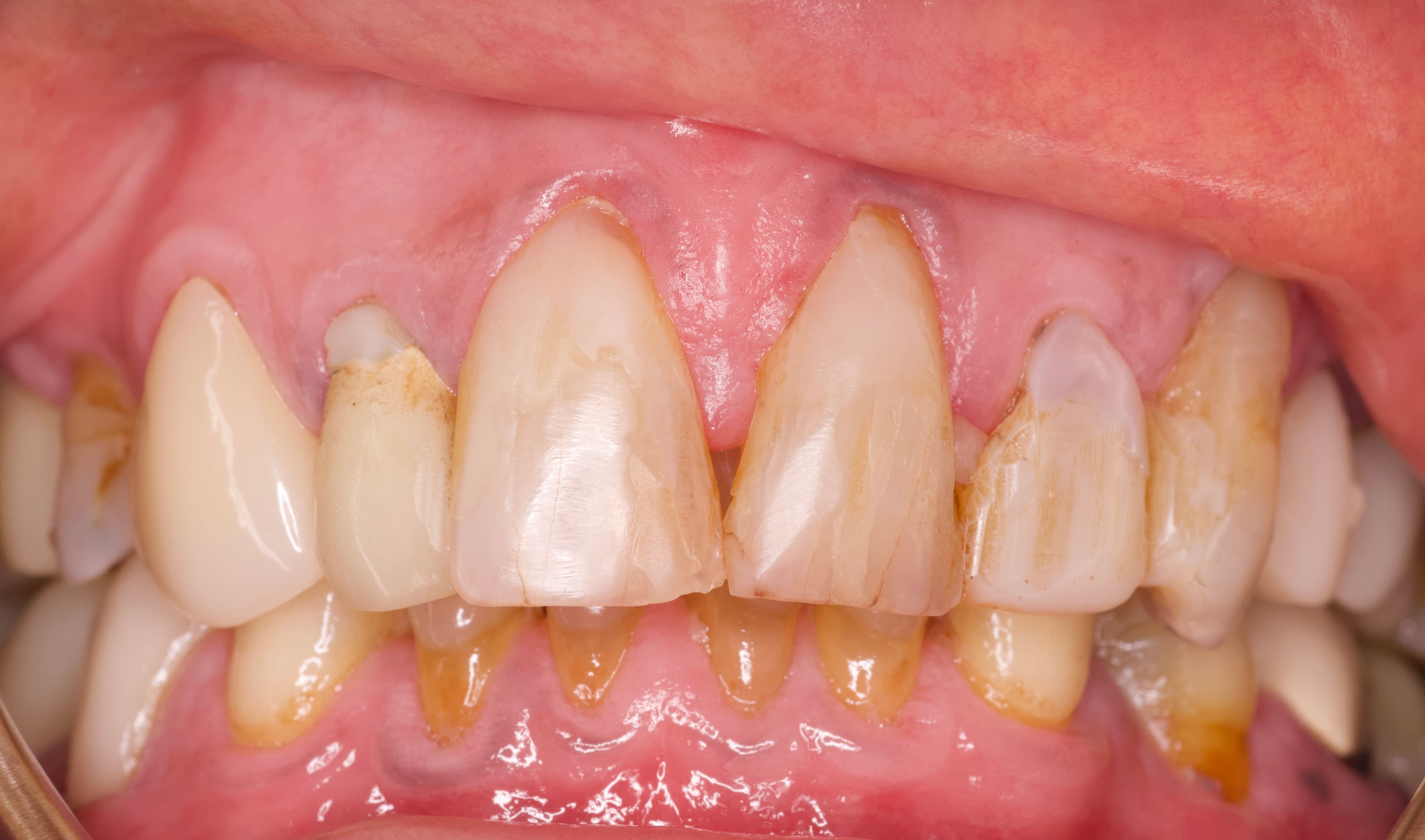
Case 01: Anterior eMax Crowns
Fifteen years of composite patch-ups had finally worn through. Conservative eMax crowns, guided by her own natural bite, restored the look and function of her original teeth.
Read the full case study →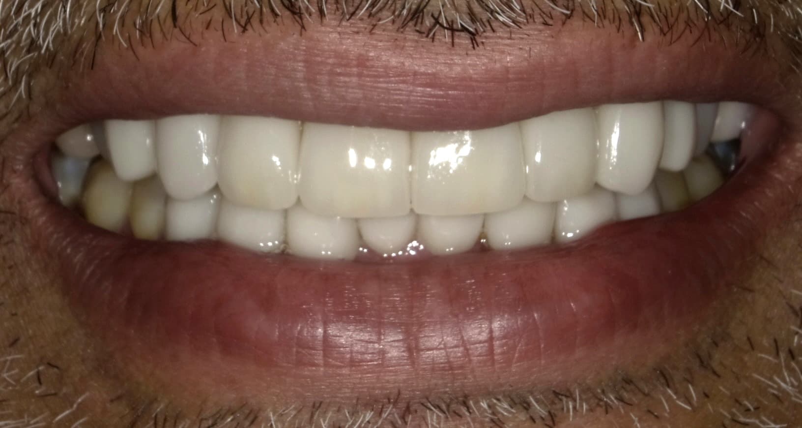
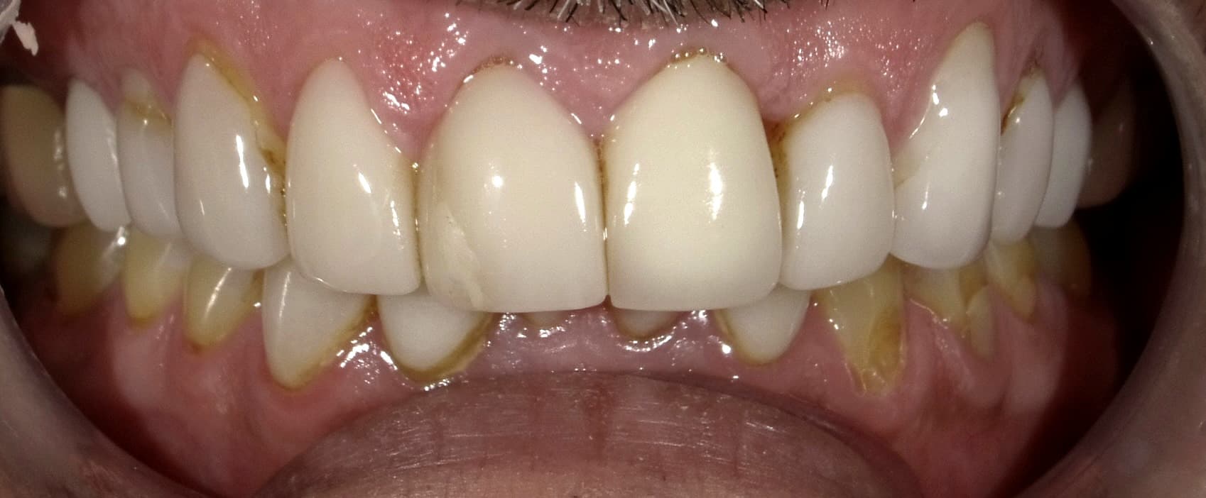
Case 02: eMax Restorations (Shade B1)
Years of chipped, worn direct composite veneers had left this patient with sensitivity and an uneven smile. Full-coverage eMax crowns in a bright B1 shade restored both balance and function.
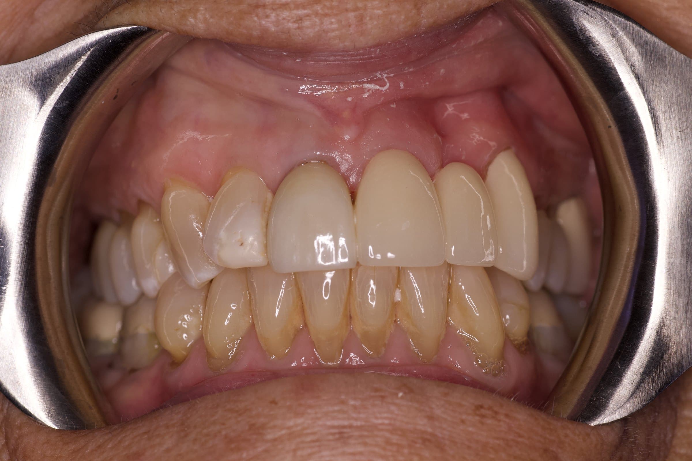
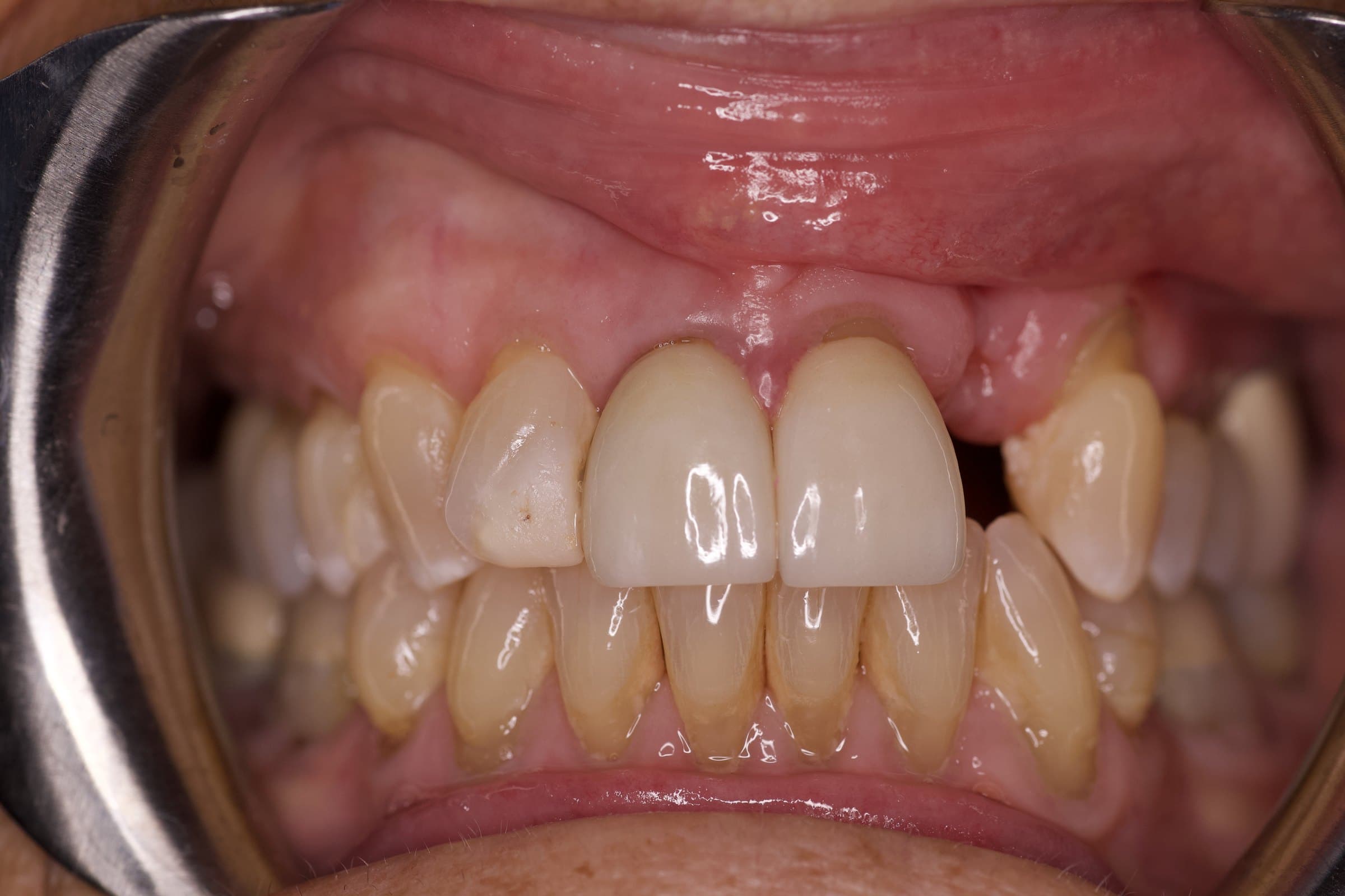
Case 03: Fixed Bridge for Missing Tooth #10
A missing front tooth had left an infection and a defect, and an implant wasn't a viable option. A porcelain-fused-to-zirconia bridge, matched to tooth #8, replaced it, and it stays in place at night.
Read the full case study →More Cases Coming Soon
The tiles below are placeholders. We do not publish a before and after until the patient has signed a release for it, so a case can sit here a while before the photographs appear in place of the tile.
Case 04: Full-Arch Implants
Case 05: Smile Makeover
Case 06: Single Implant
What you are looking at
The label on each case names the material and the tooth, not a treatment package. eMax is a ceramic used for crowns and veneers, chosen when the shade and the translucency have to sit alongside natural teeth without reading as false. A fixed bridge replaces a missing tooth by anchoring to the teeth on either side of the gap.
Tooth numbers follow the chart dentists use, counting from the back of the upper right around to the back of the lower right. Tooth #10 is the upper left lateral incisor. That is why the bridge case was photographed with the lips held back: the gum line does the talking there, and a smiling photo hides it.
How these cases are documented
The before photo is taken at the records appointment, before any work starts. The after is taken at delivery, when the final restoration goes in. Both are shot in the office, which is the only way the comparison means anything.
Retracted shots pull the lips back so you can see the gum line and the edges of each tooth. They are less flattering than a smile and far more useful. The shade and the shape are what the camera recorded.
Where a case has a written case study, the link under it walks through the diagnosis and why that restoration was chosen over the other options.
Why your case will not look like these
Each of these is one person, with one set of teeth and one history. What worked was planned around that patient's bone, their gums, their bite, and what they told us they wanted. None of it is a prediction for you, and none of it is a typical result, because there is no such thing as a typical mouth.
The honest version: we cannot tell you what is possible for your teeth from a photograph of someone else's. We can examine you, take the imaging, and give you a written plan with your options and your cost confirmed in writing at your consultation. Then you decide, with no pressure from us either way.
Cosmetic dentistry and veneers →Dental implants →Pricing and financing →
Your smile could be next